 |
|
||||||||||
|
|
 |
|
|||||||||
|
|
|
 |
|
||||||||
|
|
|
||||||||||
|
|
|
||||||||||
|
|
|
||||||||||
|
|
|
||||||||||
|
|
|
||||||||||
|
2011, Vol. 6 No. 2, Article 97
Hydatid Cyst of Muscle in Goat - A Rare Site Qazi Mudasir, K. A. Shah, Anjum Andrabi*, Qazi Nyrah1 and Muyeen A. Chesti
Central
Veterinary Hospital (Srinagar) 1Sher-e-Kashmir University of Agricultural Sciences and Technology(K), Kashmir
*Corresponding Author; e-mail address: [email protected]
ABSTRACT Hydatid cyst in the muscles is very rare and requires a high index of suspicion for its clinical and radiological diagnosis. Present communication deals with a finding of hydatid cyst with unusual location in thigh muscles in a five year old goat. The location of the lesion and therapeutic approach are discussed KEY WORDS Hydatid Cyst, muscle, goat. INTRODUCTION
Echinococcosis has been reported as one of the main health hazards all over the world. Apart from the economic damage to livestock industry it poses a serious zoonotic threat. The disease is caused by cystic larval stage of tapeworm
Echinococcus granulosus. Dog and fox are the usual hosts and serve as the main source of infection to herbivores and man as well. The infection may be contracted through ingestion/inhalation of eggs released from tapeworm segments passed in the feces by usual hosts (Aboudaya, 1985). The metacestode stage proliferates in the intermediate and aberrant hosts, predominantly in the liver by exogenous budding and by invading surrounding tissues comparable to a malignant tumour (Gottstein and Hemphill, 1997). The cyst is commonly found in liver, lungs, spleen and very rarely in muscles of intermediate host
(Sangaram and Lalitha , 2009).
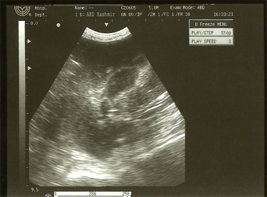
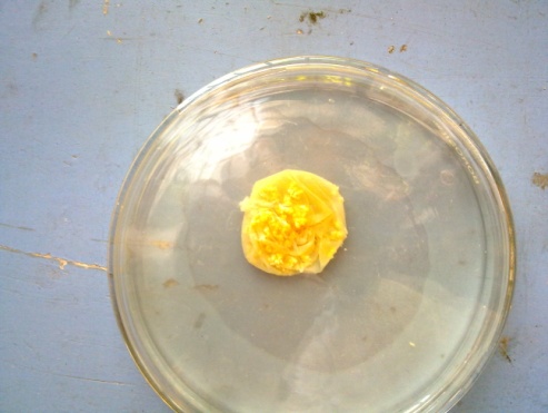
CASE HISTORY AND CLINICAL SIGNS A goat aged five years was presented at Central Veterinary Hospital, Srinagar with a history of swelling on thigh region of left leg. The swelling was not associated with symptoms like pain, dysphagia or pyrexia. The only symptom was a gradually enlarging mass on the lateral side of left thigh. There was no swelling in any other part of the body. MATERIALS AND METHODS On local examination there was a non-tender, cold and painless swelling extending above from the middle of the thigh. The swelling was smooth in surface and firm in consistency. Upper, lower medial and lateral edges were palpable. Skin overlying the swelling was free and it was not a pulsatile swelling. Ultrasonography of the affected part revealed presence of single cystic mass thick wavy in outline (Fig-1). After preparation of the site a blunt incision was given over the skin. The fascia and muscles were separated to reveal a round cystic structure lodged beneath. It was carefully separated from the surrounding areas and removed intact taking all precautions to avoid spillage of cystic contents. The cystic fluid was stained with Gentian violet. The surgical wound was closed in the routine manner and animal was administered antibiotics (Inj. Cefixime 500 mg twice daily) and analgesics (Inj Meloxicam 4 ml once daily) for next 3 days. The animal was also kept on Easy pet (Praziquintal + Pyrantal pamoate + Fenbendazole) three tablets once which was repeated after 7 days interval for 5 consecutive weeks to avoid recurrence of disease. Post-operative period was uneventful. RESULTS Incision of thigh muscle exposed a shinning white cyst wall with multiple projections at various sides (Fig-2). The inner surface of the cyst wall showed a folded appearance and displayed numerous white coloured granular structures. The proteinaceous cystic fluid revealed hydatid sand of Echinococcus granulosus on staining with Gentian violet. DISCUSSION
Echinococcus granulosus is a cyclo-zoonotic parasite and could involve human being and domestic quadrupeds like cow, sheep, goat, water buffalo, camel, pig and horse as intermediate host (Soulsby, 1982). The infection of carnivores (with immature or mature intestinal stages of
E. granulosus) does not cause morbidity, whereas the invasion of various organs (mainly liver and lungs) of intermediate or aberrant hosts by metacestode can cause severe and even fatal disease (hydatidosis). The outcome of infection in livestock and humans is cyst development in the liver, lungs, or other organ systems. In present study Hydatid cyst was detected in the thigh muscle of a goat. Hydatid cyst at different sites has been reported in goats in India (Sangaran and Lalitha, J. 2009) and abroad (Khosrow et. al.,
2011; Mohmoud and Garhy, 2002). Hydatid disease of muscle is very rare (Kacheriwala et. al., 2004 ). The lungs and liver appear to be the sites of predilection as infestation by hydatid disease most commonly occurs in the liver (55-70%) followed by the lung (18-35 %), (Guntz
et. al., 1990), (Kir and Baran, 1995) brain, heart, kidney, ureter, spleen, uterus, fallopian tube, mesentery, pancreas, diaphragm and muscles (5%) (Eshy, 1998; Singh and Dhar, 1998; Sangaram and Lalitha, 2009).
REFERENCES
FIGURES Figure-1: Ultrasonograph showing cystic cavity
Figure-2: Cyst dissected out
|
|
||||||||||
|
|
|||||||||||
|
|
|||||||||||
|
|
|||||||||||
|
|
|||||||||||
|
Copyright © Vet Scan 2005- All Right Reserved with
VetScan |
Home | e-Learning |Resources | Alumni | Forum | Picture blog | Disclaimer |
|
|||||||||
|
powered by eMedia Services |
|
||||||||||
|
|
|
|
|
|
|
|
|
|
|
|
|