 |
|
|||||||||||||||||||||||||||||
|
|
 |
|
||||||||||||||||||||||||||||
|
|
|
 |
|
|||||||||||||||||||||||||||
|
|
|
|||||||||||||||||||||||||||||
|
|
|
|||||||||||||||||||||||||||||
|
|
|
|||||||||||||||||||||||||||||
|
|
|
|||||||||||||||||||||||||||||
|
|
|
|||||||||||||||||||||||||||||
|
2010, Vol. 5 No. 2, Article 68
Management of Open Fracture and Dislocation S. Nayak*1, and S. Samantara2
1Associate Professor and Head
Department of Veterinary Surgery & Radiology,
*Corresponding Author; e-mail address: [email protected]
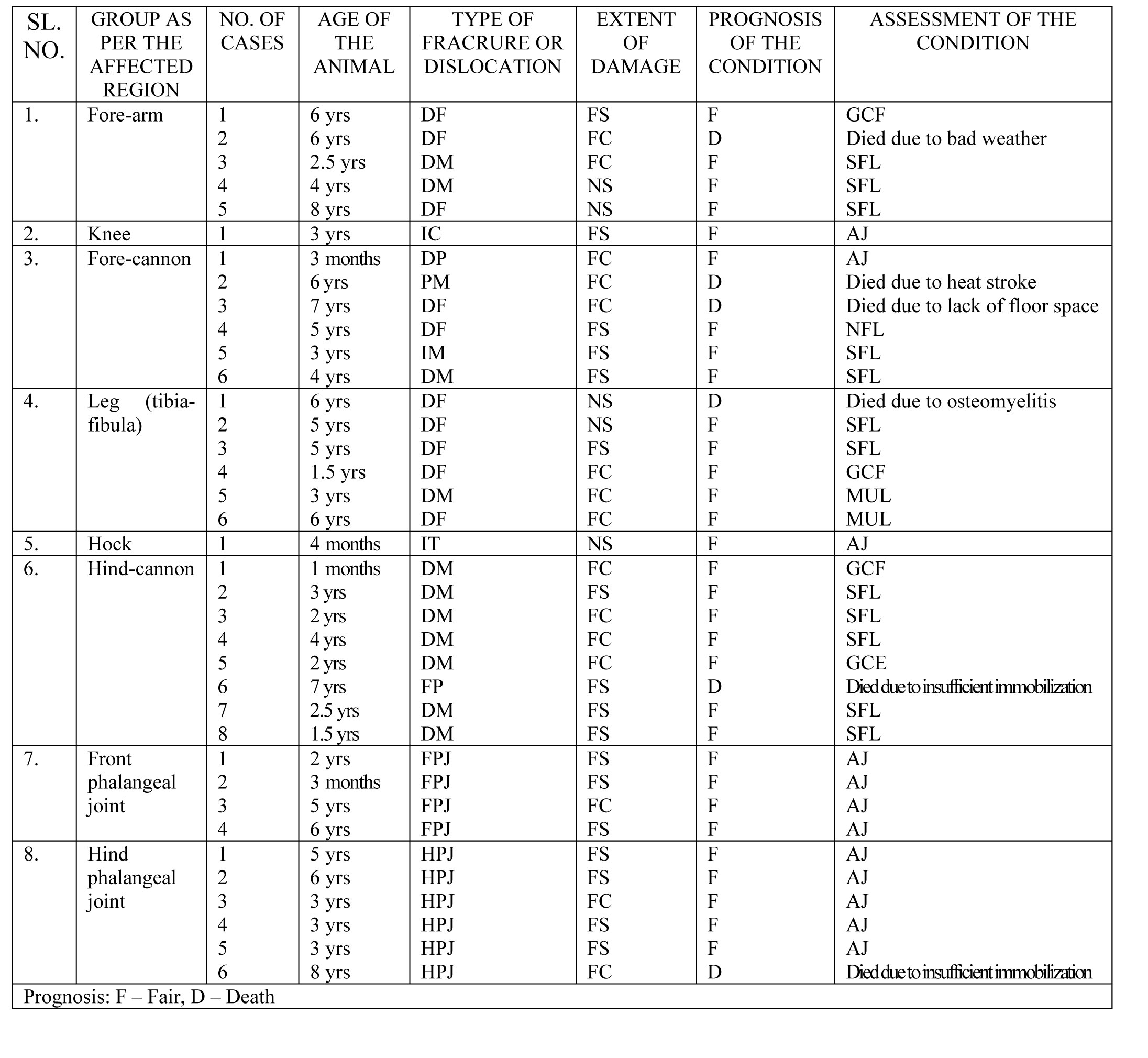
ABSTRACT Compound fracture and dislocation with open wounds in (n=37) bovines was successfully managed without amputation of the affected part by applying proper immobilizing devices, flushing with antiseptic solutions, provision of drainage facility. Proper post-operative care/nursing, regular dressing without damaging the bone marrow clot seal, provision of spacious dry shed were some of the key factors to achieve success without going for radical surgery. KEY WORDS Fracture, dislocation, bovine, amputation, radical. INTRODUCTION Fracture or dislocation which has communicating wounds piercing the skin to outside is classified under open fracture or luxation (Oehme and Prier, 1974 and Venugopalan, 1982). Due to lack of satisfactory immobilizing devices with open dressing facilities, the open fractures do not usually respond to the treatment and develop such complications that amputation remains the only measure to save the animal . Induction of arthodesis or ankylosis could be a suitable alternative approach (Stashak, 1987). During present trial an attempt was made to derive a successful surgical management of open fracture and dislocation without amputation of the affected part. MATERIALS AND METHODS Management of Compound fracture and dislocation of varying degree (Table-1) in (n=37) bovines of different breeds and age groups was achieved without amputation of the affected part. All these cases with open fractures or open dislocations at various levels of the limb were presented for treatment at the Surgery department of Orissa Veterinary College. Depending up on the time of presentation, the cases were categorized as fresh contaminated (n=15) presented within six hours of accident, fresh septic (n=17) presented within two days of occurrence and neglected cases (n=5) presented after 4-5days of occurrence. After aseptic preparation of the surrounding area the visible contaminated materials present in the wounds were flushed with warm normal saline and the left out material was remove with sterilized gauze. Then the interior of the wound was irrigated with iodine 5% lotion. The protruded bone was trimmed to blunt without disturbing the clot at the marrow cavity. Two vertical counter openings were made both at medial and lateral aspects for complete drainage of inflammatory fluid. Two soft plastic sterile catheters were introduced into the wound through the lower located openings. Both the catheters were retained in the wound at a higher level to the fracture with the help of two nylon interrupted sutures fixed to the skin. The main skin defect was closed after reducing the fracture or dislocated bone inside. The fractured part was completely covered with a thick, sterile, antiseptic padding prepared from absorbent cotton and gauze keeping both the outer ends of the catheter protruding out and bandaged. The region was kept immobilized with appropriate external immobilizers. Finally both the ends of the catheters were kept covered with sterilized gauze bandage. Additionally forearm and leg fracture was immobilized with a special type of modified Thomas splint keeping provision for dressing facilities. 10 to 15 ml of 5% iodine solution was infused through each polythene tube twice daily for seven days. The plastic catheters were removed after seven days. The animals were injected with streptopenicillin at prescribed dose rate intramuscularly for seven days. Dressing was changed at five days interval. The cases of neglected septic fractures were kept on dressing only till the dead bone pieces (sequestrum) were cast off. Open joints and dislocation cases were also dealt in the similar manner. RESULTS
Out of total thirty seven cases, only six patients died during the course of treatment. Open joint cases were cured after complete detachment of the infected synovial capsule and the dead bones (sequestrum). All the cases resulted in ankylosed joints (AJ). The animals were able to use their limbs confidently. Amongst the fracture cases, four healed forming giant callus (GCF), twelve had shortened functional limb (SFL), two had malunited limb (MUL) and only one ended in non-functional limb (NFL). DISCUSSION Rupture of the bone marrow clot at the time of debridement and open reduction contributed to the cause of osteomyelitis in most of the cases. Wound infection is the most common cause of morbidity and mortality in injured animals (Tyagi and Singh, 2002).The rate of success depends up on prompt immobilization of the part as well as control of infection (Oehme and Prier, 1974). Fresh contaminated cases with small wounds were successfully managed without sequestrum formation. Such cases healed by giant callus formation within 75 to 90 days. Infusion of iodine 5% solution in to the wounds along with simultaneous provision of drainage facility during the first week worked satisfactorily in controlling the infection .Similar observations were reported by (Tyagi and Singh, 2002). Control of infection was characterized by presence of the dry healthy scar over the original wound counter openings after 7 days. After separation of the sequestrum another 7 to 10 days of dressing and maintenance of the limbs with appropriate external immobilizers was found suitable for complete wound closure and fracture healing. The cause of death in six cases was assessed to be bad weather including heat stroke, improper care, and poor nursing during the first three days, insufficient floor space, osteomyelitis and improper immobilization of the proximal metaphyseal fracture. ACKNOWLEDGEMENTS The authors thankful to Dr. Tapan Kumar Pattanaik, Associate Professor, Veterinary Surgery and Radiology for his kind help during this research work and providing information regarding the journal Vetscan. REFERENCES
TABLES
|
|
|||||||||||||||||||||||||||||
|
|
||||||||||||||||||||||||||||||
|
|
||||||||||||||||||||||||||||||
|
|
||||||||||||||||||||||||||||||
|
|
||||||||||||||||||||||||||||||
|
Copyright © Vet Scan 2005- All Right Reserved with
VetScan |
Home | e-Learning |Resources | Alumni | Forum | Picture blog | Disclaimer |
|
||||||||||||||||||||||||||||
|
powered by eMedia Services |
|
|||||||||||||||||||||||||||||
|
|
|
|
|
|
|
|
|
|
|
|
||||||||||||||||||||