|


2009, Vol. 4 No. 2, Article 43
Ultrasonographic Diagnosis of the Bovine
Genital Tract Disorders
Vijay Kumar and G. N. Purohit
Department of Animal Reproduction, Gynaecology & Obstetrics
College of Veterinary & Animal Science, Bikaner
*Corresponding Author;
e-mail address: [email protected]
ABSTRACT
Cows brought to the clinics (85) for pregnancy diagonosis or treatment of various reproductive problems were examined by transrectal ultrasonography (TRUS). The various problems diagnosed were endometritis (23), post-partum metritis (6), pyometra (13), mucometra (7), follicular cyst (17), luteal cyst (5), abortion (2), mummified foetus (3), macerated foetus (1), adhesions (1), anestrus (6) and ovarian abscess (1). indicating that TRUS can be used efficiently for diagnosing reproductive disorders and the response to treatment thereafter.
KEY
WORDS
TRUS, Genital tract disorder.
INTRODUCTION
In bovine practice, ultrasonography has become an important diagnostic tool for evaluating the female reproductive system. Its importance in diagnosis lies in the non-invasiveness of the instrument (Carriere et. al., 2002). In spite of its immense use in supplementing diagnosis during physiological states, its use in delineating different pathological conditions of the bovine genital tract continues to be less frequently described. This study was conducted on clinical cases presented to the veterinary gynaecology and obstetrics outdoor to record the sonographic appearance during different pathological conditions of genital tract.
MATERIALS AND METHODS
Cows presented to the veterinary gynaecology and obstetrics outdoor (n=85) with different pathological conditions were included in the present study. Transrectal sonographic examination was performed using a portable ultrasound machine (AGROSCAN linear, ECM 1"6 BD de la Republique, F 16000 Angouleme, FRANCE), with a linear array dual frequency probe (5.0/7.5 MHz). The images were saved in a multimedia kit attached to the instrument and subsequently
transferred to the computer. Animals were examined by rectogenital palpation and ultrasonography was done later to confirm / potentiate the clinical diagnosis. After administration of an appropriate treatment sonography was done again to determine the effect of treatment.
RESULTS
Among a total of 85 clinical cases investigated during present study, the different clinical conditions diagnosed were endometritis (23), post-partum metritis (6), pyometra (13), mucometra (7), follicular cyst (17), luteal cyst (5), abortion (2), mummified foetus (3), macerated foetus (1), adhesions (1), anestrus (6) and ovarian abscess(1).
(Table 1)
DISCUSSIONS
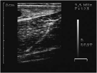
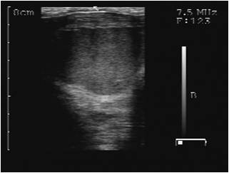
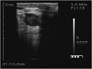
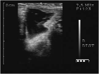
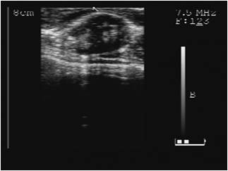
The presence of anechoic fluid in cases of endometritis along with snowy echogenic particles observed during the present study (Fig. 1,2) was similar to previous reports of (Fissore et al. 1986; Kasaimanickam et al. 2004; Lenz et al. 2007; Barlunda et al. 2008). Amongst the various pathological conditions diagnosed during the present study endometritis was the most frequently diagnosed pathology. that caused repeat breeding in dairy cows as these cases were brought to the clinics with complaint of repeat breeding with an apparently normal cervico-vaginal discharge. This finding corroborated previous reports of (Miller et al. 1980; Le Blanc et al. 2002).
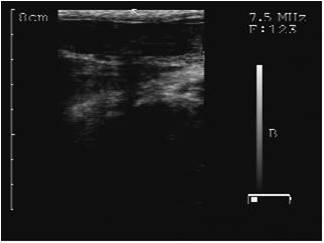
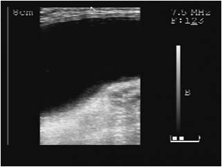
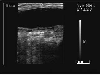
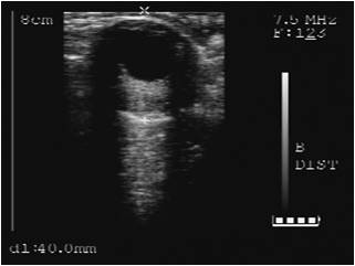
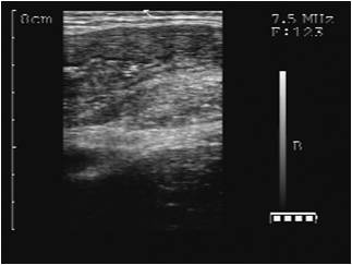
The sonographic features of post partum metritis were in part similar to endometritis; however, there was enormous amount of anechoic fluid in the distended uterus alongwith echogenic particles (Fig.3). falling in line with features reported by (Fissore et al., 1986; Melendez et al., 2004). Subsequent to therapy, the uterine diameter, fluid accumulation and echogenic particles got reduced (Fig.4).
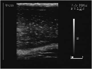
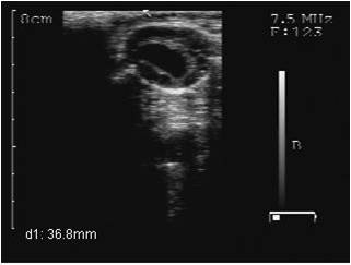
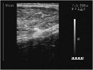
In cases of pyometra, sonographically, echogenic pus inside the uterus was observed (Fig.5). Most of these cases were in fact presented for pregnancy diagnosis. Ultrasonographic appearance confirmed the diagnosis of pyometra. Diagnosis of pyometra by ultrasonography had previously been done on the basis of high volume of accumulated echogenic uterine content without fetus and cotyledons, closed cervix and corpus luteum on the ovary (Bon Durant, 1999; Sheldon and Dobson, 2004; Sheldon et al., 2006; Fissore et al., 1986). treatment of pyometric cows was instituted resulting into discharge of huge quantity of pus (Fig.6).
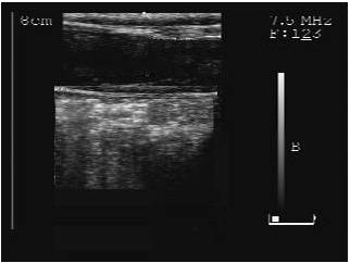
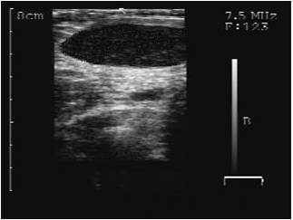
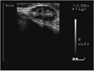
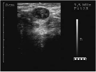
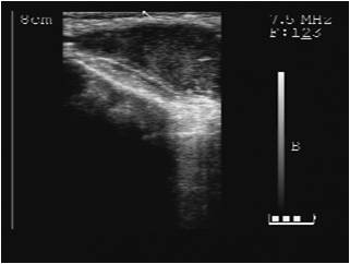
In cases of mucometra, a consistent ultrasonographic finding was thin walled uterus which was filled with anechoic fluid (Fig.7,8) similar to a observations of (Fissore et al., 1986. outl of 22 cases of ovarian cyst evaluated during the present study, 77.28% (17/22) were follicular cysts (Fig. 9) and 22.72% (5/22) were luteal cysts (Fig.11). A higher incidence of follicular cysts has been recorded by (Gaur and Purohit, 2007; Durocher et al., 2005). The sonographic difference between the two types of cysts was the appearance of distinct hypoechoic wall (2.5 mm) of luteal tissue in luteal cysts. The diameters of ovarian cysts varied from 30mm to 48mm. The proportion of cows that showed multiple follicular cysts was only 29.41% (5/17) of the total follicular cysts diagnosed. The sonographic features of cysts were simple fluid filled (anechoic) thin walled structures ³ 25mm (follicular cysts) or round to ovoid structures ³ 25mm with a thick wall and centrally an anechoic area falling in agreement with findings of (Fissore et al., 1986; Kuber and Jalakas, 2002; Farin et al. 1992; Ribadu et al., 1994; Farin et al., 1990; Naoki, 2007). previously ,Farin et al., 1990 consider sector scanning better for diagnosis of luteal cysts however,during the present study ovarian cysts could easily be diagnosed by linear array scanning (Kuber and Jalakas, 2002; Ribadu et al., 1994; Farin et al., 1992; Naoki, 2007) also used linear array scanning for such cases. The response to the treatment was good as evident by the sonographic disappearance of both the type of cysts (Fig.10,12) within 10-40 days of treatment, conforming previous reports of (Walton et al., 1990; Morales-Roura et al., 1998; Yavas and Walton, 2000). The diagnosis of single ovarian cysts is always found difficult by rectal palpation alone, but can easily be confirmed by ultrasonography. Farin et al., 1992 had previously recorded a sensitivity and specificity of 43.3% and 64.7%respectively for diagnosis of ovarian cysts by rectal palpation whereas it was 86.7% and 82.3% by ultrasonography In the cases of anestrus the sonographic findings were a persistent corpus luteum (Fig.13) in 33.3% (2/6) of the cases, or poor follicular growth in 66.67% (4/6) of the cases during the present study. The sensitivity and specificity of ultrasonography in diagnosing ovarian structures has been shown to be 95 and 100% respectively (Ribadu et al. 1994).
The sonogram also proved to be useful supplement to confirm abnormalities like mummified fetus, macerated fetus, adhesion, abortion (Fig.14-17) and ovarian abscess (Fig. 18). In conclusion ultrasonography of the bovine uterus and ovaries can help in definitive diagnosis of various pathological conditions.
REFERENCES
-
Barlunda C.S, Carrnthersa T.D, Waldnera C.L and Palmer C.W (2008), A comparision of diagnostic techniques for postpartum endometritis in dairy cattle. Theriogenol. 69:714-723.
-
BonDurant, RH (1986). Examination of the Reproductive Tract of the Cow and Heifer. In: Current Therapy in Theriogenology: Diag., Treat. and Prev. of Rep. Dis. in Ani., edn 2, pp 95-101.
-
Brown E.M, Elmore R.G, Garverick H.A (1982). Gonadotropin releasing hormone treatment of dairy cows with ovarian cysts II. Histology of ovarian cyst walls. Theriogenol. 17, 689-696.
-
Calder MD, Salfen BE, Bao B, Youngquist RS, Garverick HA. (1999) Administration of progesterone to cows with ovarian follicular cysts results in a reduction in mean LH and LH pulse frequency and initiates ovulatory follicular growth. J. Ani. Sci. 77:3037-42.
-
Carrière PD, DesCôteaux L, Durocher J.(2002). Evaluation échographique du tractus reproducteur bovin : Développement normal et anormal des follicules ovariens et du corps jaune. Méd Vét du Québec, 32 :128-131.
-
Durocher J, Morin N, Blondin P.(2005). Effect of hormonal stimulation on bovine follicular response and oocyte developmental competence in a commercial operation. Theriogenol., 65:102-115.
-
Farin PW, Youngquist RS, Parfet JR, Garverick HA. (1990). Diagnosis of luteal and follicular ovarian cysts in dairy cows by sector scan ultrasonography. Theriogenol. 34: 633-42.
-
Farin PW, Youngquist RS, Parfet JR, Garverick HA. (1992). Diagnosis of luteal and follicular ovarian cysts by palpation per rectum and linear-array ultrasonography in dairy cows. J Am Vet Med Assoc. 200:1085-9.
-
Fissore RA, Edmondson AJ, Pashen RL and Bondurant RH (1986) The use of ultrasonography for the study of the bovine reproductive tract II non pregnant, pregnant and pathological conditions of the uterus. Ani. Rep. Sci. 12, 167-177.
-
Garverick T.S(1997). Ovarian follicular cysts in dairy cows. J. Dairy Sci. 80,995-1004.
Gaur, M. and G. N. Purohit (2007). Follicular dynamics in Rathi (Bos indicus) cattle. Vet. arhiv 77, 177-186.
-
Kasimanickam R, Duffield TF, Foster RA, Gartley CJ, Leslie KE, Walton JS, Johnson WH (2004). Endometrial cytology and ultrasonography for the detection of subclinical endometritis in postpartum dairy cows. Theriogenol., 62:9-23.
-
Kübar H, Jalakas M. (2002). Pathological changes in the reproductive organs of cows and heifers culled because of infertility. J Vet Med A Physiol Pathol Clin Med. 49:365-72.
-
Le-Blanc SJ, Duffield TF, Leslie KE, Batemann KG, Keefe GP, Walton JS(2002). Defining and diagnosing postpartum clinical endometritis and its impact on reproductive performance of dairy cows. J Dairy Sci.;85:2223-36.
-
Lenz M, Drillich M, Heuwieser W. (2007). Evaluation of the diagnosis of subclinical endometritis in dairy cattle using ultrasound. Berl Munch Tiera. Woch.. 120:237-44.
-
Melendez P, McHale J, Bartolome J, Archbald LF, Donovan GA (2004). Uterine involution and fertility of Holstein cows subsequent to early postpartum PGF2alpha treatment for acute puerperal metritis. J Dairy Sci.;87:3238-46.
-
Miller HV, Kimsey PB, Kendrick JW, Darien B, Doering L, Franti C. (1980) Endometritis of dairy cattle: diagnosis, treatment and fertility. Bov Pract; 32:13-23.
-
Morales-Roura JS, Hernandez-Ceron J, Vazquez-Garcia JA.( 1998) Effect of HCG treatment at the time of insemination on luteal function and fertility in repeat-breeding Holstein cows. Veterinaria, Mexico;29:269-72.
-
Naoki S.B .(2007). Follicular cysts in dairy cows. Ani. Sci J.;78:1-6.
Peter AT.(2004) An update on cystic ovarian degeneration in cattle. Reproduction in Domestic Animals;39:1-7.
-
Ribadu AY, Ward WR, Dobson H.(1994). Comparative evaluation of ovarian structures in cattle by palpation per rectum, ultrasonography and plasma progesterone concentration. Vet Rec.135:452-7.
-
Sheldon IM, Dobson H.(2004) Postpartum uterine health in cattle. Ani. Rep. Sci.;82/83:295–306.
-
Sheldon IM, Lewis G, LeBlanc S, Gilbert R.(2006) Defining postpartum uterine disease in dairy cattle. Theriogenol.;65:1516–30.
-
Walton JS, Gary WH, Nancy AR, Kenneth EL. (1990) Effects of progesterone and human chorionic gonadotrophin administration five days post insemination on plasma and milk concentrations of progesterone and pregnancy rates of normal and repeat breeder dairy cows. Canadian Journal of Veterinary Research; 54:305-8.
-
Wiltbank MC, Gümen A, Sartori R.( 2002). Physiological classification of anovulatory conditions in cattle. Theriogenol;57:21-52.
-
Yavas Y, Walton JS.(2000) Induction of ovulation in postpartum suckled beef cows: a review . Theriogenol; 54:1-23.
Table 1: Transrectal ultrasonographic findings in various reproductive problems
|
Type of case
|
No. of animals (n)
|
Ultrasonographic
finding
|
|
Endometritis
|
23
|
Moderate to large amount of anechoic fluid in lumen
alongth with "snowy" echogenic particles. Uterine
wall is thickened.
|
|
Post-partum Metritis
|
6
|
Uterine wall is thickened and uterine body is
distended. Enormous amount of anechoic fluid is
present in the lumen along with with echogenic
particles.
|
|
Pyometra
|
13
|
Uterus distended and have thickened uterine wall.
The viscous fluid inside the lumen contained
diffused, echogenic particles, floating in it.
Sonographic image resembles with uterine tissue.
|
|
Mucometra
|
7
|
Uterus is thin walled which is visible as white
streak and appears to be filled with echogenic
particles in the enormous anechoic fluid present
inside it.
|
|
Follicular cyst
|
17
|
Imaged as thin walled (≤2mm) non-echogenic
structures visible over the surface of the ovary.
Size is
≥25 mm
|
|
Luteal cyst
|
5
|
Anechoic central cavity bordered by a distinct wall
(2-5 mm) of luteinized tissue imaged over the
ovarian surface. Size >25 mm.
|
|
Abortion
|
2
|
Hypoechogenic membrane loosed their tense appearance
and begins to appear as floating with in the
anechoic uterine lumen. The conceptus, with
membranes may be imaged in any area of birth canal
accompanied by anechoic fluid. CL generally
maintains good size and echogenicity for a number of
days post abortion.
|
|
Macerated fetus
|
1
|
Uterine wall were thickened and bone fragments were
seen as dense echogenic areas shadowing the tissue
below. Surrounding and between the bones purulent
fluid was present with its typical appearance of a
non echogenic background containing many small
echogenic particles.
|
|
Mummified foetus
|
3
|
Uterine fluids were not evident in contrast to the
snowy appearance of the lumen of uteri in which
macerated foetus were seen. Uterine wall is
thickened. Fetal parts are rarely identified and
mostly appeared as a poorly defined echogenic mass.
|
|
Adhesions
|
1
|
Medium or thick band of hypoechoic streak is
observed along the uterine wall.
|
|
Anestrus
|
6
|
In 2 of the cases CL was imaged
as distinctly echogenic areas with in the ovarian
stroma while in rest of the cases, on sonographic
scanning, no echogenic area was imaged in the
ovarian stroma and small follicles (2-5 mm diameter)
was visible on the ovarian surface.
|
|
Ovarian abscess
|
1
|
A pus filled cavity is observed over the ovary
imaged as fine dots of echogenic particles in the
anechoic fluid matrix.
|
|
 |