 |
|
||||||||||
|
|
 |
|
|||||||||
|
|
|
 |
|
||||||||
|
|
|
||||||||||
|
|
|
||||||||||
|
|
|
||||||||||
|
|
|
||||||||||
|
|
|
||||||||||
|
2009, Vol. 4 No. 1, Article 35
Lacrimal apparatus of Iranian river Buffaloes A S Bighama* and M Shadkhastb
*Corresponding Author; e-mail address: [email protected]
ABSTRACT The gross anatomy of the nasolacrimal duct of ten buffalos (Bubalus bubalis) was studied. Anatomic casts were obtained after infusion of corrosion cast (Rodopas cast) material. The findings were similar to that of cattle described earlier. The present study would be useful for identification of congenital and clinical affections involving the lacrimal system of buffaloes. KEY WORDS Nasolacrimal duct, gross anatomical study, buffalo. INTRODUCTION Buffalo rearing has great economical importance in the Asian agriculture. They are an important source of draft power, milk, meat, and hide. As per current estimates, 150 million Asian buffaloes produce 77 million tons of milk and 3 million tons of meat. In several countries they also contribute up to 30% of the draft power for agricultural operations (10). Information on nasolacrimal apparatus system in buffaloes is limited. The lacrimal apparatus provides a passage for tear drainage from the eye to the nasal cavity. The system for each eye in most species consists of dorsal and ventral lacrimal puncta, paired canaliculi, lacrimal sac and the nasolacrimal duct (11). The nasolacrimal system of cattle has been studied in detail(6,15). Dacrocystorhinography, the radiographic visualization of the lacrimal apparatus using radiographic contrast media, has been used to study normal anatomy (4,14) as well as pathologic conditions of the nasolacrimal duct in human beings (7), dogs (3), horses (8), sheep (4), cattle (6,15), camels (14), cats (4) and llamas (9,13). The present study describes normal anatomical course of lacrimal duct in buffaloes and would be useful for identification of congenital or clinical affections. MATERIALS AND METHODS Ten adult buffalo heads (separated proximal to the third cervical vertebra) were collected from a local slaughterhouse. On gross observation, the dorsal and ventral puncta were located in the medial canthus area of eyelids. They were sufficiently wide in diameter and could be catheterized without much difficulty (Fig 1 and 2). The distal opening of nasolacrimal duct was found in nasal cavity and was catheterized with a 4-F tube (Fig 3). Casting material was infused in retrograde direction. 4-6 milliliter of casting material was required to fill the entire length of naso lacrimal duct. The casting material was allowed to solidify. Nasal cavity was exposed from lateral side by removing segments of the lacrimal, zygomatic, maxillary & incisive bones. The lacrimal apparatus was then examined and its details recorded. Visualization of the medial side was facilitated by removal of the ethmoturbinates, ventral conchae and lacrimal bone. RESULTS On gross examination the lacrimal puncta appeared slit-like openings, 0.5-1.0 mm in diameter, 4.0-10.0 mm away from the medial canthus located 1.0 mm from the mucocutaneous junction of the palpebral margin. The paired ventral and dorsal canaliculi were 7 to 9 mm long and converged into a small dilatation, the lacrimal sac located in the orbit on the fossa of the lacrimal bone. The nasolacrimal duct extended from the lacrimal sac to the nostril in the wall of the nasal cavity (fig 4). The proximal portion of the lacrimal duct (75 to 80 mm in length) was in the osseous lacrimal canal. The average total length of lacrimal duct was 260 mm. The osseous lacrimal canal with a slight curve at its origin was directed rostrally. It passed the lacrimal, zygomatic, and maxillary bones lateral to the lacrimal sinus before moving dorsal to the maxillary sinus. The middle portion of the nasolacrimal duct passed through the osseous lacrimal canal after crossing the ventral conchal crest. The duct covered only by nasal mucosa and a thin connective tissue membrane then traversed the nasal cavity in a curved (descending) fashion. The distal opening of the nasolacrimal duct was 1.5 to 2 mm in diameter located on the medial surface of the lateral nasal wall about 40 mm above the dorsal angle of the nostril. DISCUSSION The details of the nasolacrimal system of various domestic animal species have already been described (4,5,13,1,2,12). The findings of this study revealed the details of lacrimal apparatus in buffaloes. The lacrimal apparatus consisted of an orbital part and a nasal part. The orbital lacrimal apparatus consisted of a simple lacrimal sac and paired canaliculi with the dorsal and ventral puncta. All findings were similar to those of cattle (6,15). In pigs only one punctum and a duct are present (11). In dogs the punctum and the duct of the ventral eyelid is occasionally absent (4). The nasolacrimal duct coursed rostrally in a curved fashion. This finding was similar to that of cattle(6,15).However in llamas the nasolacrimal duct courses rostrally in a sigmoid fashion (13). The nasolacrimal duct in the present study involving buffaloes was uniform in diameter throughout its length unlike horses (8). ACKNOWLEDGMENT The financial support rendered by Faculty of Veterinary Medicine of Shahre-kord University is greatly appreciated. REFERENCES
Fig. 1: Dorsal(a) and Ventral(b) puncta 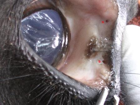 Fig. 2: Ventral puncta catheterization 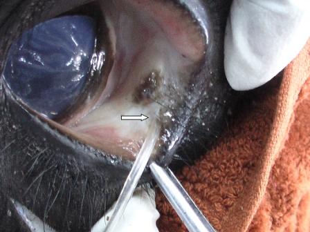 Fig. 3: Distal opening of nasolacrimal duct catheterization 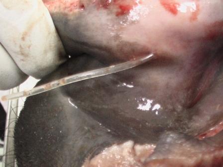 Fig. 4: Proximal part of lacrimal duct on the lateral surface of nasal cavity 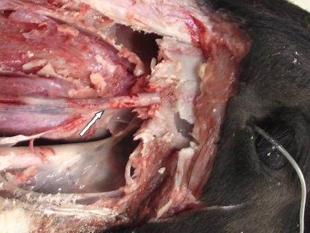
|
|
||||||||||
|
|
|||||||||||
|
|
|||||||||||
|
|
|||||||||||
|
|
|||||||||||
|
Copyright © Vet Scan 2005- All Right Reserved with
VetScan |
Home | e-Learning |Resources | Alumni | Forum | Picture blog | Disclaimer |
|
|||||||||
|
powered by eMedia Services |
|
||||||||||
|
|
|
|
|
|
|
|
|
|
|
|
|