|


2010, Vol. 5 No. 1, Article 55
Chemotheraputic Management of Canine Mammary Tumors
Ankur Sharma*, M. S. Dhakate and S. V. Upadhye
Department of Veterinary Surgery and Radiology,
Nagpur Veterinary College,
Maharashtra Animal and Fishery Sciences University,
Seminary Hills, Nagpur, Maharashtra (INDIA) 440006
*Corresponding Author;
e-mail address: [email protected]
ABSTRACT
A study on treatment of mammary tumor in bitches was conducted by comparing two regimens of chemotherapy viz. Oncotron (Group-I, n=6) and a combination of Oncotron and Mammofen (Group-II, n=6). Comparative analysis of the two groups revealed significant variations in haematobiochemical values. Regression percentage in Group I was 66.20 percent and in Group II was 69.56 percent with recurrence observed in one case in each group. The G-banded karyotype of lymphocyte in metaphase obtained showed normalcy with respect to number, length and breadth of the chromosomes with normal appearance of bands.
KEY
WORDS
Mammary Tumor, Karyotyping, Oncotron, Mammofen.
INTRODUCTION
Mammary gland tumors represent 25-50 percent of all the neoplasms in bitches (Moulton et al, 1970). Classical modalities of cancer therapy include surgery, radiation and chemotherapy. Chemotherapy yields best results when used to treat rapidly growing tumors and is beneficial in dogs having multiple negative prognostic factors and lymph node metastasis. Drugs like Doxorubicin, Mitoxantrone, Cisplatin, Vinblastine, Cyclophosphamide, etc. may be indicated in dogs with aggressive tumors. Anti-estrogens like- Tamoxifen, Toremifene, etc. are used as adjuvant therapy after surgery or in combination with chemotherapy, which reduces the chances of hormone receptors positive breast cancer recurrence.
Consequently the present study was conducted to see the efficacy of intravenously administered Mitoxantrone alone and in combination with orally administered Tamoxifen.
MATERIALS AND METHODS
Twelve number of bitches with mammary tumors (less than 3 cm dia.) were randomly divided in two groups
of six each. Group-I animals were treated with chemotherapeutic agent Mitoxantrone (Oncotron) alone @ 5mg/m2 by intravenous route on a scheduled interval of 0, 21st and 42nd day and Group-II animals were treated with Mitoxantrone supplemented with Tamoxifen tablet (Mammofen) @10mg B.I.D orally daily for three months. Comparative analysis of both the groups was carried out with reference to clinical examination, haemato-biochemical alterations, recurrences or metastasis
(if any) and karyotyping, recorded at weekly intervals up to 49th day.
The cytogenetic study aimed with identification of neoplasia associated chromosomal aberration in canine mammary tumor was carried out as per the method specified by Moorehead
(1960), where 20 metaphase cells each from lymphocytes of 12 bitches suffering with mammary tumor were identified and compared with the karyotype of lymphocytes of clinically normal bitch.
RESULTS AND DISCUSSION
The clinical parameters of animals under both the treatment regimes fluctuated within normal range throughout the period of observation.
Haematology:
The haematological parameters (Table-1) revealed significant reduction in Total Erythrocyte Count (TEC), Total Leucocyte Count (TLC) and Total Platelet count (TPC) in both the groups. Significant neutropenia, lymphocytosis and monocytosis were also observed. Haemoglobin (Hb) values were reduced but not significantly. Mitoxantrone chemotherapy induced myelosuppression as cytotoxic drugs suppress the replicating precursor cells of the bone marrow and attributed to significant haematological changes. These finding were in accord with a previous reports of Dobson and Gorman,
(1993) and Pavesi et al, (1995).
Biochemical Parameters:
Significant elevation of biochemical values were observed in both the groups after administration of Mitoxantrone. However, the increase in the values was within the normal physiological range (Table 2). The alterations in biochemical observations were in accordance with earlier reports (Benjamin, 1979; Brar et al, 2002) who observed that chemotherapeutic drugs alone or in combination with tamoxifen were hepatotoxic and elevated biochemical values.
Regression:
The use of Mitoxantrone alone in Group I showed a regression percentage of 66.20 percent and
Mitoxantrone and Tamoxifen in Group II were found to be moderately effective with success rate of 69.56 percent. However, recurrence was observed in one case in both the groups. Similar results were reported in different studies (Conzen et al, 1966; Fisher et al, 2001; Moore et al, 1994 and Ogilvie et al, 1991) who observed a partial or complete remission in
Mitoxantrone treated patients to the extent of 41-56 % with no significant difference of
Tamoxifen regimen over chemotherapy alone.
Chemotherapy with Mitoxantrone was found to be safe and moderately effective therapy. Common side effects observed due to chemotherapeutic treatment included anaemia, lethargy, nausea, vomiting and mild alopecia.
Lymphocyte Culture Technique and Karyotyping:
The short-term blood culture technique which was employed gave good culture. The karyotyping findings were in agreement with earlier studies (Jayshree et al, 1994; Patel, 1995). The lymphocyte karyotype picture of normal bitch indicated that thirty-eight pairs of chromosomes were acrocentric whereas the pair of sex chromosomes was metacentric (2n = 78). Chromosome No. 1 and X chromosome were found to be the largest chromosomes and the length of rest of the chromosomes was in decreasing order. The chromosome numbers recorded in the bitches affected with mammary tumor during present trial were similar to that of lymphocyte of clinically normal bitch. Genetic involvement could not be proved only on the basis of gross study of chromosomes and it was felt that the study at gene or molecular level is required to be undertaken.
The response to tumor was unaffected by presence of anti estrogen status, i.e. supplementation of Tamoxifen showing no significant advantage over that achieved from chemotherapy alone.
REFERENCES
-
Benjamin, M.M., 1979. Haematology,In:Outline of Veterinary Clinical Pathology.3rd Edn.Iowa State Univ. Press, Ames, Iowa. pp 51-56.
-
Brar, R.S., H.S. Sandhu and Singh, A., 2002. Veterinary Clinical Diagnosis by Laboratory Methods. 1st edn. Kalyani Publishers, New Delhi.
-
Conzen, S. D., P.A. Kaufman, C. Arvizu, P. LeMarbre, L.H. Maurer, L.A. Mott and L.E. Mills, , 1996. Phase II trial of tamoxifen, etoposide, mitoxantrone and cisplatin (TEMC) in patients with metastatic breast carcinoma. 78(9):1906-11.
-
Dobson, J.M. and Gorman, N.T, , 1993. Cancer Chemotherapy in Small Animal Practice. Blackwell Scientific Publication, London. 67-154.
-
Fisher, B., Anderson, S., Tan-Chiu, E., Wolmark, N., Wickerham, D.L., Fisher, E.R., Dimitrov, N.V, Atkins, J.N., Abramson, N., Merajver, S., Romond, E.H., Kardinal, C.G., Shibata, H.R., Margolese, R.G. and Farrar, W.B, 2001. Tamoxifen and Chemotherapy for Axillary Node-Negative, Estrogen Receptor – Negative Breast Cancer: Findings from National Surgical Adjuvant Breast and Bowel Project B-23. Journal of Clinical Oncology, 19(4): 931-942.
-
Jabara, A.G, 1969. Two cases of mammary neoplasms arising in male dogs. Aust. Vet. J., 45: 476-480.
-
Jayshree, R.; P. Thangraju, A.M. Nainar and A.R. Krishnan: Karyological studies in dogs. Indian Anim Sci. 69(3):183-184.
-
Moore, A.S., Ogilive, G.K., Ruslander, D., Rand, W.S., S.M. Cotter, Getzy, D.M., L’Heureux, D.A. and Dennis, R.A, 1994. Evaluation of mitoxantrone
for the treatment of lymphoma in dogs. J Am Vet Med Assoc.
204(12):1903-5.
-
Moorehead, P.S, 1960. Chromosomal preparations of leucocytes cultured from human peripheral blood. Expt. Cell Res. 20:613-616.
-
Moulton, J.E.; D.O.N. Taylor, Dorn, C.R. and Anderson, A.C., 1970.: Canine mammary tumor. Vet. Pathology., 7: 289-320.
-
Ogilvie, G.K.; Obradovich, J.E., Elmsli, R.E., Vail, D.M., Moore, A.S., Straw, R.C., Dickinson, K., Cooper, M.F. and Withrow, S.J., 1991. Efficacy of mitoxantrone against various neoplasms in dogs. J Am Vet Med Assoc. 198(9):1618-21.
-
Patel, R.K.; U. Radhakrishna and V.K. Khoda, 1995. A modified technique of GTG banding in cattle and buffaloes. Nucleus, 38:37-39.
-
Pavesi, L.; Preti, P.G., Da Prada Pedrazzoli, P., Poggi, G. and Robustelli della, C.G., , 1995. Epirubicin versus mitoxantrone in combination chemotherapy for metastatic breast cancer. Anticancer Res. 15 (2) :495-501.
Table-1 Effect of Chemotherapy on Haematological
Parameters (click to enlarge)
Table-2 Effect of Chemotherapy on Biochemical Parameters
(click to enlarge)
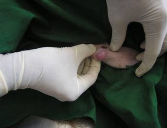
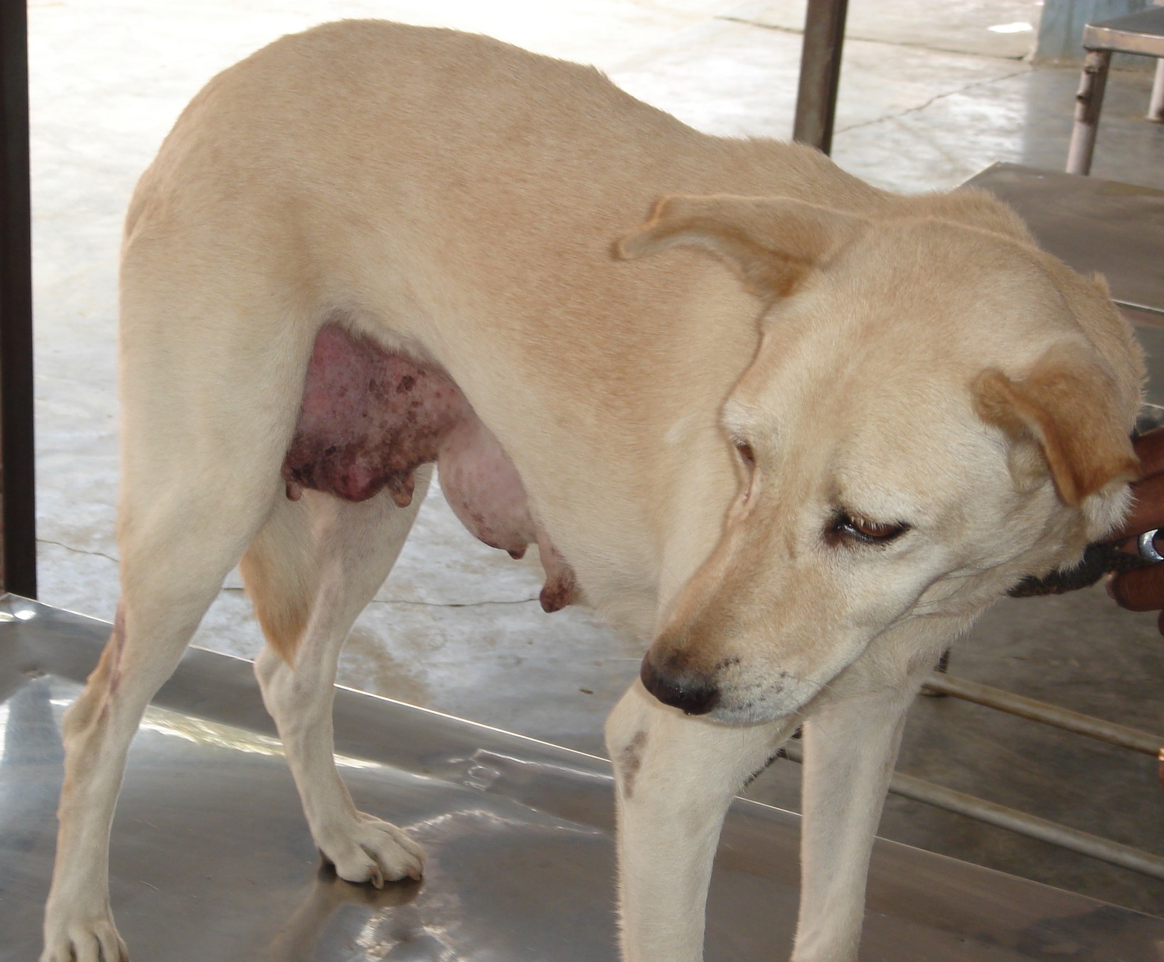
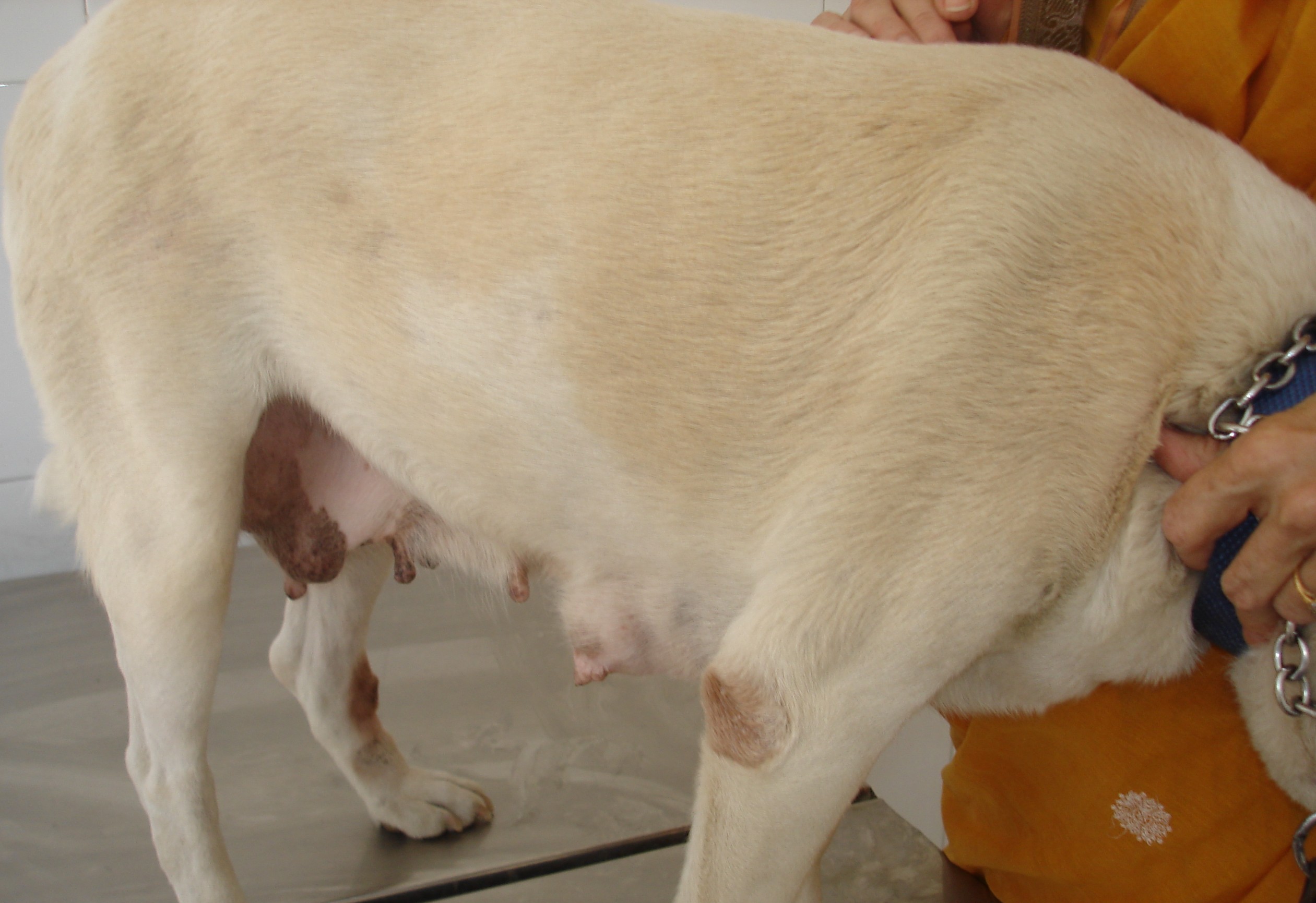
Group- I Mitoxantrone Chemotherapy
|
Fig 1:
Mammary tumor on right caudal abdominal mammary gland
on day ‘0’. |
Fig 2: Moderate
regression in mammary tumor growth on ‘49th’ day. |
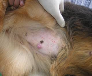
Group-II Mitoxantrone And Tamoxifen Therapy
|
Fig 3: Mammary tumor growth on right caudal abdominal and
inguinal mammary glands on day 0. |
Fig 4:
Regression in the tumor size on post treatment day 49th. |
G-Banded Karyotype of a Peripheral Lymphocyte In Metaphase
(click to enlarge)
 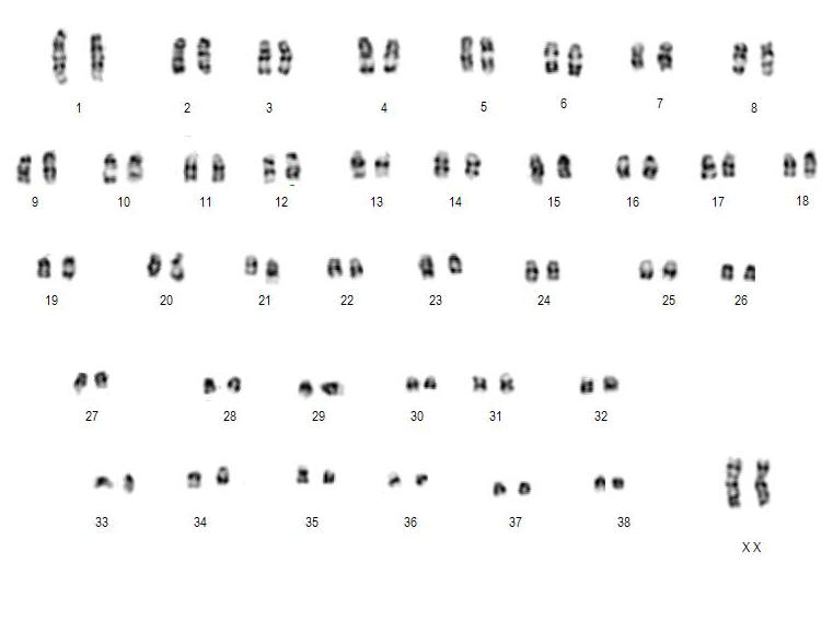
| Fig 5: Non-descript female dog affected with mammary tumor |
Fig 6: Normal non-descript female dog |
|
 |